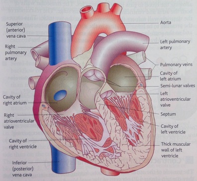
40 structure of the heart with labels
A Beginner's Guide to the Hearts Structure | Heart Surgery Information The hearts structure is divided into four main parts called chambers. These four chambers are divided into two groups: The Atriums are located within the upper part of the heart. There is one is on the right side, and the other is on the left side. Oxygenated blood is pumped through the left atrium into the body. Diagrams, quizzes and worksheets of the heart | Kenhub Worksheet showing unlabelled heart diagrams. Using our unlabeled heart diagrams, you can challenge yourself to identify the individual parts of the heart as indicated by the arrows and fill-in-the-blank spaces. This exercise will help you to identify your weak spots, so you'll know which heart structures you need to spend more time studying ...
How the Heart Works: Diagram, Anatomy, Blood Flow The heart is an amazing organ. It starts beating about 22 days after conception and continuously pumps oxygenated red blood cells and nutrient-rich blood and other compounds like platelets throughout your body to sustain the life of your organs.; Its pumping power also pushes blood through organs like the lungs to remove waste products like CO2.; This fist-sized powerhouse beats (expands and ...

Structure of the heart with labels
Diagram of Human Heart and Blood Circulation in It Four Chambers of the Heart and Blood Circulation. The shape of the human heart is like an upside-down pear, weighing between 7-15 ounces, and is little larger than the size of the fist. It is located between the lungs, in the middle of the chest, behind and slightly to the left of the breast bone. The heart, one of the most significant organs ... posterior view of heart labeled - scissorsandcombtattoos Heart right lateral view The heart is a muscular organ that pumps blood around the body by circulating it through the circulatoryvascular system. Anterior View Of Human Heart Anatomy is a photograph by Alayna Guza which was uploaded on September 7th 2017. Outside view of the back posterior of the heart. Start studying Heart Anatomy Posterior View. Draw And Label The Heart - Draw A Labelled Diagram Of The Human Heart ... Draw And Label The Heart - Draw A Labelled Diagram Of The Human Heart Two atria and two ventricles and couple of blood vessels opening into them. Click here to get an answer to your question ️ draw a diagram of the human heart and label its parts. Diagram of the human heart.
Structure of the heart with labels. Correctly Label The Following External Anatomy Of The Anterior Heart The right atrium, the anterior descending aorta, and the vasculature are all essential organs of the cardiovascular system. The external anatomy of the human heart consists of the four chambers that form the apex of the heart. Each chamber has an apex that corresponds to a box. There are two boxes on each side of the heart: the atria and the ... Heart - Pathology Outlines - Histology Newly oxygenated blood returns to the left atrium via 4 pulmonic veins at a normal pressure of approximately 10 mmHg. Left atrial contraction moves blood through the mitral valve into the left ventricle. Pacemaker of the heart located near the sinotubular junction, where the superior vena cava meets the right atrium. Drag the labels to identify structural components of the heart. labeling Activity: The Sectional Anatomy of the Heart (Part 2) Drag the labels to identify structural components of the heart. Roe Interventricular septum Aortic arch Moderator band Cusp of mitral Valve Chordae tendinea ATRIUM Intertrial seplum Cusp... Free Heart Worksheets for Human Anatomy Lessons Print out sheet of the human heart with labels - This fun heart worksheet shows kids the different parts of the heart. They'll learn about the left ventricle, the left atrium, the tricuspid valve, and more. Human Heart Clipart - There is a coloring page, heart labeling worksheet and heart anatomy chart. Clipart is a fun way for kids to ...
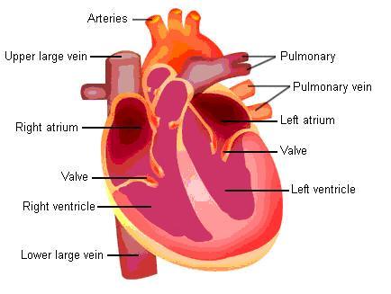
Heart: Anatomy | Concise Medical Knowledge - Lecturio Heart: Anatomy. The heart is a 4-chambered muscular pump made primarily of cardiac muscle tissue. The heart is divided into 4 chambers: 2 upper chambers for receiving blood from the great vessels, known as the right and left atria, and 2 stronger lower chambers, known as the right and left ventricles, which pump blood throughout the body. Heart: illustrated anatomy - e-Anatomy - IMAIOS This interactive atlas of human heart anatomy is based on medical illustrations and cadaver photography. The user can show or hide the anatomical labels which provide a useful tool to create illustrations perfectly adapted for teaching. Anatomy of the heart: anatomical illustrations and structures, 3D model and photographs of dissection. Structure Of Heart Diagram Without Labelling - File Diagram Of The ... The heart is made up of two chambers: Heart, organ that serves as a pump to circulate the blood. Diagram_of_the_human_heart_(no_labels).jpg (608 × 600 pixels, . Without labels to quiz yourself on the main cardiac structures and . "can you label this heart diagram, and identify key blood flow through the heart. Without labels to quiz ... Anatomy, Thorax, Heart - StatPearls - NCBI Bookshelf The heart is a muscular organ situated in the center of the chest behind the sternum. It consists of four chambers: the two upper chambers are called the right and left atria, and the two lower chambers are called the right and left ventricles. The right atrium and ventricle together are often called the right heart, and the left atrium and left ventricle together functionally form the left ...
Anatomy of the heart and coronary arteries (coronary CT) - IMAIOS Anatomy of the human heart and coronaries: how to view anatomical structures. This tool provides access to an MDCT atlas in the 4 usual planes, allowing the user to interactively discover the heart anatomy. The images are labeled, providing an important medical and anatomical tool. The quiz mode makes it possible to evaluate the user's progress. PDF Label The Heart Diagram Answers Bookmark File PDF Label The Heart Diagram Answers(Heart Quiz) [IGCSE/GCSE] Heart Structure - Memorize In 5 Minutes Or Less! Blood Flow through the Heart in 2 MINUTES Heart and major blood vessels quiz.Download e copies of my text books from campbellteaching.co.uk Cardiovascular System 1, Heart, Structure and Function Labeling The Heart Heart ... Know the Structures and Functions about Your Heart Here are some more information about heart structure and function: The human heart is just roughly about the size of a fist. Your heart weighs about 10 - 12 ounces (or 280 - 340 grams) if you are a man, and 8 -10 ounces (or 230 - 280 grams) if you are a woman. In an adult, the heart beats at an average of 60-80 times per minute. The newborn's ... Correctly Label The Following Internal Anatomy Of The Heart The aorta, or aortic arch, is the outermost layer of the heart. The left ventricle is covered with the ventricular aorta, and the pulmonary veins are located inside the aorta. The two atria, the left and right aorta, and the right aortic arch are all external organs. These organs carry oxygen-rich blood to the body.
Heart-final exam - Anatomy & Physiology 1121 with Breeding at Sinclair Community College - StudyBlue
Heart - Wikipedia The heart has four chambers, two upper atria, the receiving chambers, and two lower ventricles, the discharging chambers.The atria open into the ventricles via the atrioventricular valves, present in the atrioventricular septum.This distinction is visible also on the surface of the heart as the coronary sulcus. There is an ear-shaped structure in the upper right atrium called the right atrial ...

Human Heart Pictures with Labels Best Of File Diagram Of the Human Heart Hug Wikimedia Mons ...
Understanding an ECG | ECG Interpretation | Geeky Medics An ECG electrode is a conductive pad that is attached to the skin to record electrical activity. An ECG lead is a graphical representation of the heart's electrical activity which is calculated by analysing data from several ECG electrodes. A 12-lead ECG records 12 leads, producing 12 separate graphs on a piece of ECG paper.
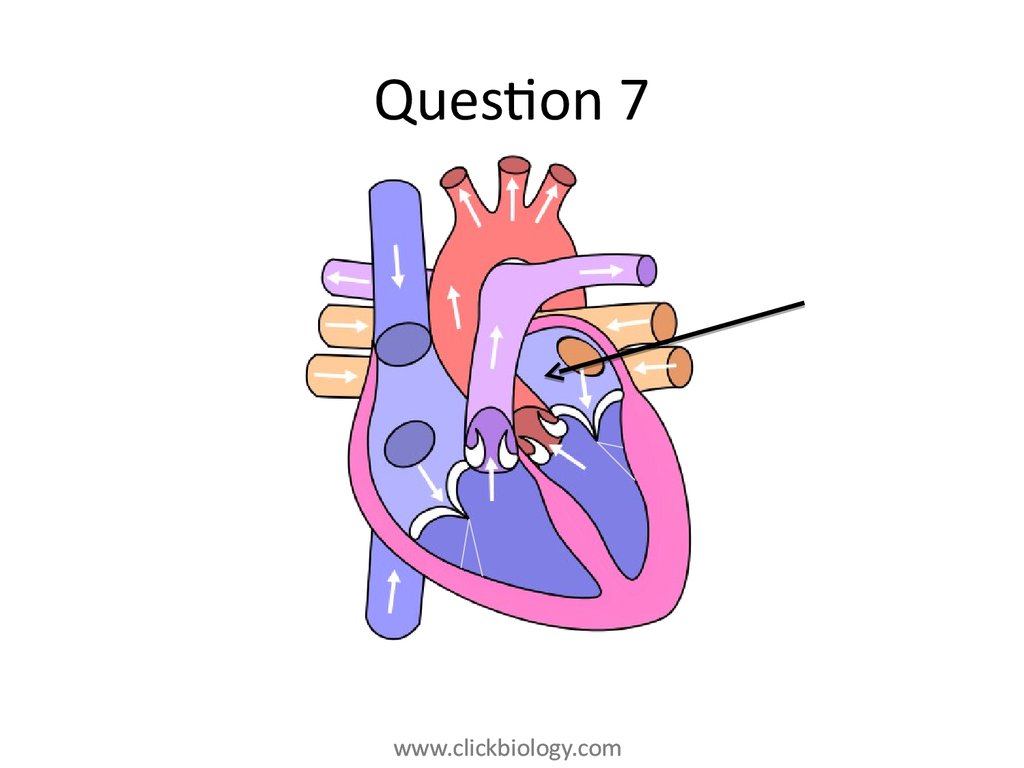
Heart structure and function - online presentation
Diagram Labeled Of The Heart : Label The Heart Quiz : Labeled diagram ... The top panel shows the human heart with the arteries and veins labeled. Learn all about the anatomy and physiology of the human heart with an interactive diagram and detailed descriptions of the organ and its . 8 human heart structure chalkboard art posters anatomy diagram cardiologist gift. Diagram of circulatory system with main parts labeled.

Simplified Heart Labeled Decal | Shop Fathead Anatomical Images Graphics
Structure And Label Of The Heart / Anatomy Of A Human Heart This page discusses the heart anatomy. The heart is a muscular organ about the size of a fist, located just behind and slightly left of the breastbone. Png, jpg, gif · no labels version · azərbaycanca · català · english · english · hrvatski · italiano · lingua franca nova. Your heart is the main organ of your circulatory system.

Abdominal Anatomy Male - Human Body Diagram Without Labels , Transparent Cartoon, Free Cliparts ...
Reinforcement: Anatomy of the Human Heart Reinforcement: Anatomy of the Human Heart. I created this worksheet for my anatomy students to review the anatomy of the heart. Students focus on vocabulary that relates to anatomical features, such as the mitral valve, aorta, and heart chambers. At the end, students label a line drawing of the heart. I included a word bank at the top, but you ...

The human egg cell explained for egg donors | Altrui
Heart Labeling Quiz: How Much You Know About Heart Labeling? Here is a Heart labeling quiz for you. The human heart is a vital organ for every human. The more healthy your heart is, the longer the chances you have of surviving, so you better take care of it. Take the following quiz to know how much you know about your heart. Questions and Answers. 1.
Know the Structures and Functions about Your Heart | New Health Advisor
Heart anatomy: Structure, valves, coronary vessels | Kenhub Heart anatomy. The heart has five surfaces: base (posterior), diaphragmatic (inferior), sternocostal (anterior), and left and right pulmonary surfaces. It also has several margins: right, left, superior, and inferior: The right margin is the small section of the right atrium that extends between the superior and inferior vena cava .
![Untitled Document [www.bio.sunyorange.edu]](http://www.bio.sunyorange.edu/updated2/THINKING_EVOLUTION/physiology1/heart/c_chambers.jpg)
Untitled Document [www.bio.sunyorange.edu]
Interior With View Heart Of The Labels Human Heart Diagram Anatomy Picture Valves Arteries Human heart diagram, anatomy, picture, valves & arteries. 4 2014, carolina biological supply company mammalian heart dissection answer sheet internal observations of the heart insert your photograph interior with view heart of the labels of the interior view of the heart. label each of the structures listed in activity 1, step 4 of your lab ...

Attack of The Silverfish!: Images of the stinging cells of Cnidaria
Parts of the Heart & Blood Flow | Diagram & Overview - Study.com The four parts of the heart are the right atrium, right ventricle, left atrium, and left ventricle. The atrium receives blood from the body, the right ventricle pumps blood to the lungs, the left ...
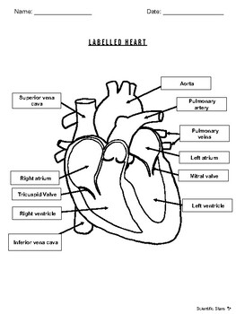
Labelled Heart by Scientific Stars | Teachers Pay Teachers
How the Heart Works - The Heart | NHLBI, NIH The Heart. The heart is an organ about the size of your fist that pumps blood through your body. It is made up of multiple layers of tissue. Your heart is at the center of your circulatory system. This system is a network of blood vessels, such as arteries, veins, and capillaries, that carries blood to and from all areas of your body.
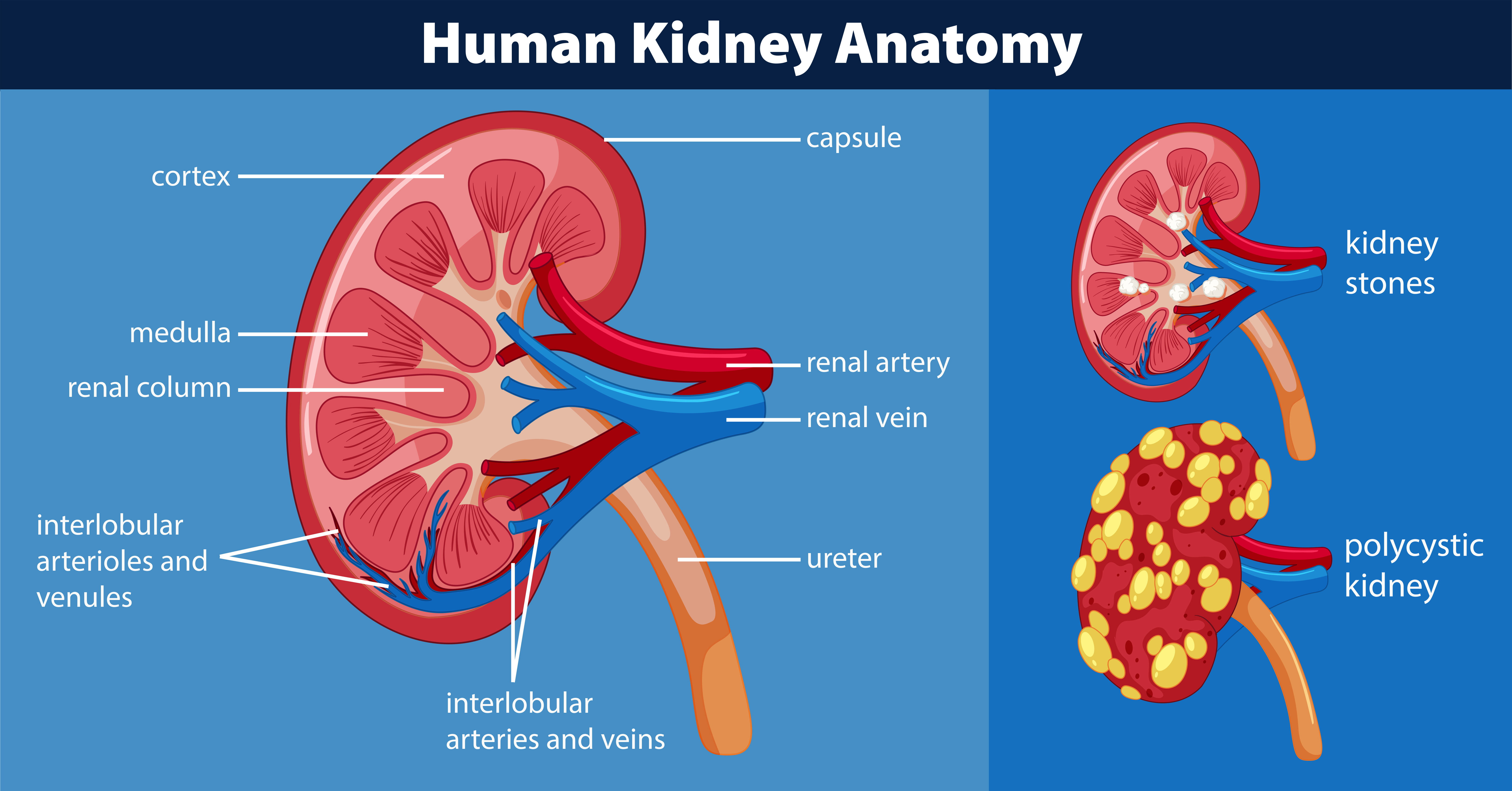
Human kidney anatomy diagram 446409 Vector Art at Vecteezy
Draw And Label The Heart - Draw A Labelled Diagram Of The Human Heart ... Draw And Label The Heart - Draw A Labelled Diagram Of The Human Heart Two atria and two ventricles and couple of blood vessels opening into them. Click here to get an answer to your question ️ draw a diagram of the human heart and label its parts. Diagram of the human heart.

Labelled Heart by abpischools - Teaching Resources - Tes
posterior view of heart labeled - scissorsandcombtattoos Heart right lateral view The heart is a muscular organ that pumps blood around the body by circulating it through the circulatoryvascular system. Anterior View Of Human Heart Anatomy is a photograph by Alayna Guza which was uploaded on September 7th 2017. Outside view of the back posterior of the heart. Start studying Heart Anatomy Posterior View.
5.1 The Structure of the Heart - AQA A-Level Biology Revision
Diagram of Human Heart and Blood Circulation in It Four Chambers of the Heart and Blood Circulation. The shape of the human heart is like an upside-down pear, weighing between 7-15 ounces, and is little larger than the size of the fist. It is located between the lungs, in the middle of the chest, behind and slightly to the left of the breast bone. The heart, one of the most significant organs ...
Tissues Flashcards | Easy Notecards

Parts Of Heart Diagram Stock Illustration - Download Image Now - iStock
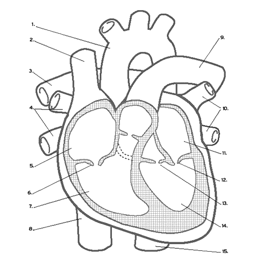
Heart Labeling (Internal)
Post a Comment for "40 structure of the heart with labels"